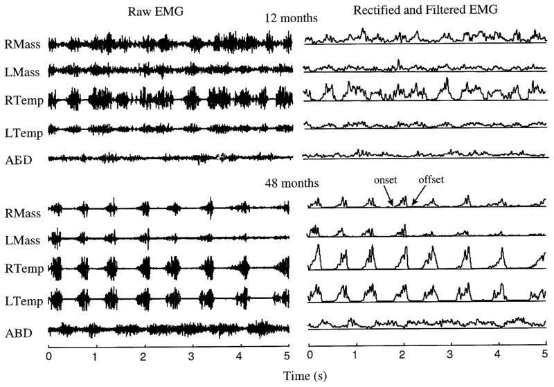
FIG. 1.
Electromyographic (EMG) activity during chewing by the same subject at 12 and 48 mo of age. Left: raw EMG activity. Right: rectified and filtered signals. Modulation of the EMG signals at 12 mo is much more poorly defined than in the later samples. RMass, right masseter; LMass, left masseter; RTemp, right temporalis; LTemp, left temporalis; ABD, anterior belly of digastric.

