
Fig. 2. SEM microphotographs of SPCL scaffolds.
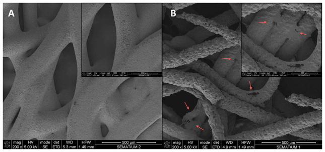
A – SPCL scaffold alone; B – SPCL scaffold loaded with ASCs (magnifications of each image on upper right corners). Red arrows pointing some of the ASCs spread on SPCL fibers.
Official websites use .gov
A
.gov website belongs to an official
government organization in the United States.
Secure .gov websites use HTTPS
A lock (
) or https:// means you've safely
connected to the .gov website. Share sensitive
information only on official, secure websites.
A – SPCL scaffold alone; B – SPCL scaffold loaded with ASCs (magnifications of each image on upper right corners). Red arrows pointing some of the ASCs spread on SPCL fibers.