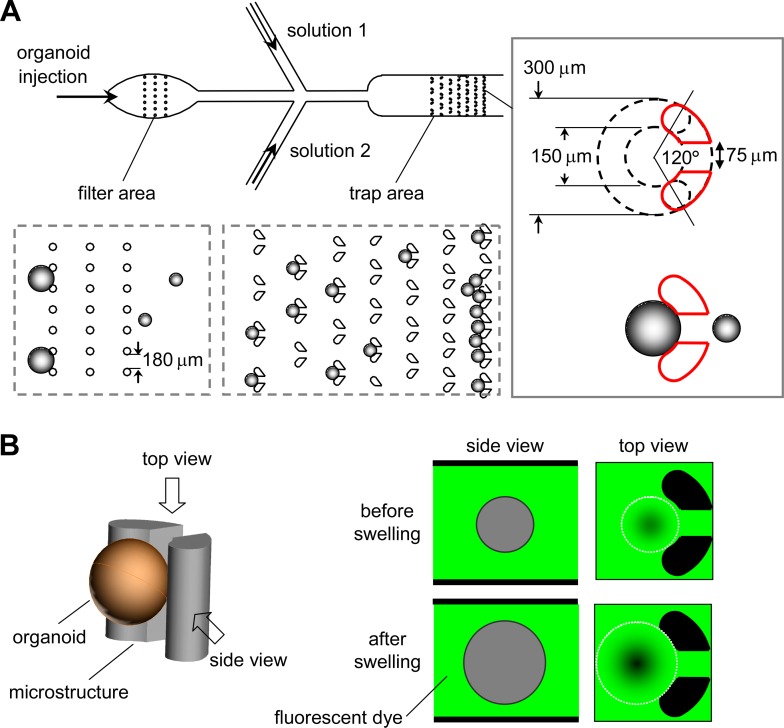
Figure 1.
Microfluidic channel design and volume measurement principle. (a) Channel design, showing passage of an enteroid suspension through a filter region to remove large particles and debris, and then into a trap region to immobilize for microscopy. Side injection ports allow continuous perfusion with different solutions. Inset at right shows post-pair geometry. (b) Principle of enteroid volume determination by fluorescent dye exclusion in which the perfusion solution contains a membrane-impermeant fluorescent dextran. Enteroid-excluded volume reduces area-integrated fluorescence signal.

