
Figure 3.
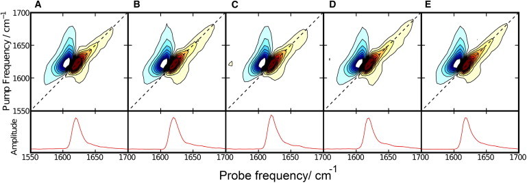
Two-dimensional infrared spectroscopy confirms that all peptides form amyloid fibrils. (A) hIAPP. (B) H18L-hIAPP. (C) H18Q-hIAPP. (D) Free CT-hIAPP. (E) H18Q free CT-IAPP. The diagonal slices of each spectrum are shown at the bottom of the figure. To see this figure in color, go online.