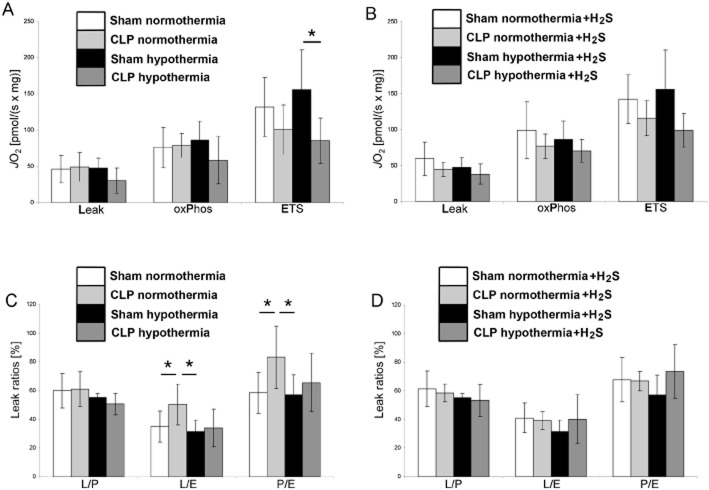
Figure 1.
Effect of H2S and hypothermia on mitochondrial respiration in mechanically permeabilized small murine liver biopsies. Eight groups of anaesthetized and mechanically ventilated mice were studied in order to combine all the treatments of interest, namely sham operation versus CLP (closed white and black bars vs. light and grey bars), normothermia (38°C, white and light grey bars) versus hypothermia (27°C, black and dark grey bars) and i.v. application of the sulfide donor Na2S (diagrams B and D) or placebo (diagrams A and C). The study design as well as the methods used to measure mitochondrial respiratory activity in the tissue samples is described in Baumgart et al. (2010). Briefly, mitochondrial respiration was measured in terms of oxygen flux (JO2) per mg wet tissue under maximum stimulation by complex I + II substrates and ADP in the coupled (OxPhos) and FCCP-induced uncoupled (ETS) conditions. The Leak state (Leak) was obtained in the OxPhos state by inhibiting the ATP synthesis by oligomycin, and represents the respiratory activity necessary to compensate for the proton leakage, slipping and cations exchange along the inner mitochondrial membrane. Relating the Leak state to the OxPhos state and to the ETS state yields the L/P and L/E ratios, respectively, the P/E ratio is the ratio OxPhos to ETS state (see diagrams C and D). Data are shown as mean ± SEM of n = 6 determinations, *P < 0.05.

