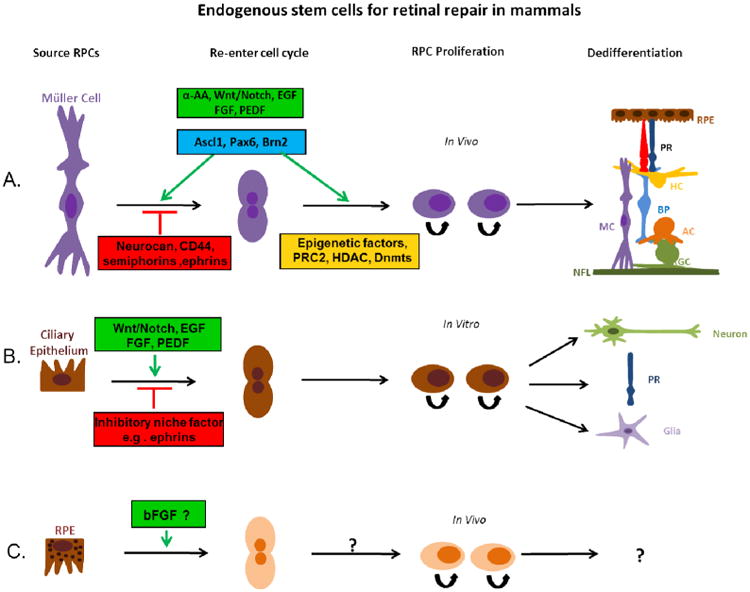
Figure 2.

Müller cells, retinal pigment epithelium and Ciliary epithelium cells are reported as retinal stem cells for retinal repair in mammals. (A) Latent Müller cells are stimulated or suppressed to re-enter the cell cycle by different factors (green or red boxes). Müller cells can be induced to proliferate by growth factors and transcription factors listed in the green boxes and differentiate into various retinal cell types. Modification of epigenetic factors may also contribute to the regulation of the stem cell potency of Müller cells. (B) It has been shown that in vitro, ciliary epithelium cells isolated from mature retina can be stimulated (green box) or suppressed (red box) to re-enter the cell cycle, differentiating into new neurons, including photoreceptors (PR), and glia cells. (C) Retinal pigment epithelium has also been shown to possess the ability to proliferate and transdifferentiate into retinal neurons, but the neurogenic potential of these cells in mammals are fairly restricted and appear to be deficient in the regulatory elements that are required for induction of transdifferentiation. EGF: Epidermal Growth Factor; FGF: Fibroblast Growth Factor; PEDF: Pigment Epithelium-Derived Factor; α-AA: α-Aminoadipic Acid; Ascl1: Achaete-Scute Complex-like 1; Pax6: Paired Box Gene 6; Brn2: Brain-2; PRC2: polycomb repressive complex-2; HDAC: Dnmts: DNA methyltransferases; RPC: Retinal Progenitor cell; PR: Photoreceptor; RPE: Retinal Pigment Epithelium; HC: Horizontal cell; BP: Bipolar cell; MC: Müller cell; AC: Amacrine cell; RGC: Retinal Ganglion cell; NFL: Nerve Fiber layer
