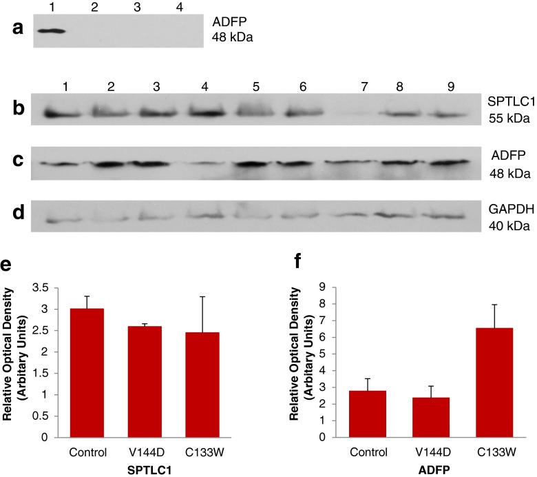
Fig. 3.
Expression of lipid droplet marker protein ADFP in HSN-1 patient-derived lymphoblasts. a Immunoblot detection of ADFP in oleic acid-treated and oleic acid-untreated total cell lysates; 1 represents treated controls; 2, untreated controls; 3, untreated V144D mutant; and 4, untreated C133W mutant. Immunoblots of total protein lysates from oleic acid-treated cells probed for SPTLC1 (b), ADFP (c), and GAPDH (d); 1–3 represents treated controls; 4–6, treated V144D mutants; and 7–9, treated C133W mutants. Histograms of the immunoblotting results of oleic acid-treated controls and of SPTLC1 (e) and ADFP (f), normalised to GAPDH (n = 3)

