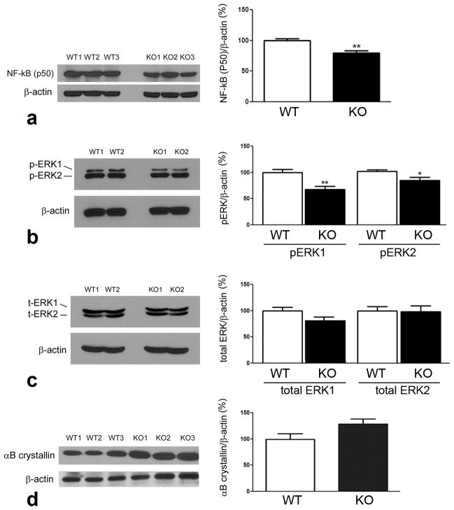
Fig. 6. Western blot analysis of NFκB (p50), ERK and αB crystallin proteins.
Neural retinas were harvested from σR1+/+ (WT) and σR1−/− (KO) mice, protein isolated, subjected to SDS-PAGE followed by immunoblotting to detect (a) NFκB (p50), (b) phosphorylated ERK1 and ERK2, (c) total ERK1 and ERK2, (d) αB crystallin. Band densities were normalized to β-actin. Densitometric analysis of the bands normalized by β-actin are provided adjacent to each set of blots. (n = 4–5 mice per group; *p<0.05,**p<0.01)

