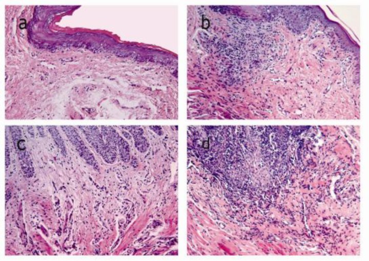
Figure 2.
Histological compared to a: normal appearance in the negative (Original magnification, ×200).Subepithelial connective tissue including fibroblasts, endothelial cells, gingival epithelial cells and few inflammatory cells b: Mild inflammation histological appearance in the prophylactic group. c: Moderate inflammation histological appearance in the therapeutic group. d: Severe inflammation histological appearance in the positive group

