Figure 1.

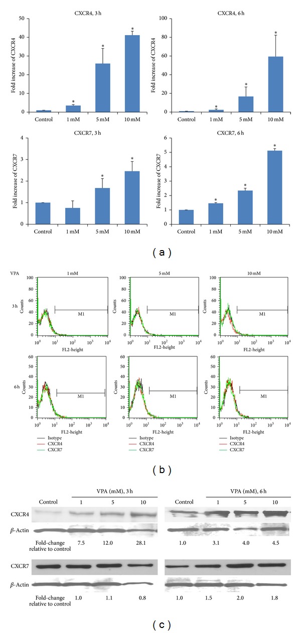
VPA increases CXCR4 and CXCR7 gene and protein expression. CB-derived MSC were treated with 0 (control), 1, 5, or 10 mM of VPA for 3 or 6 h. (a) Expression of CXCR4 and CXCR7 mRNAs was evaluated by real-time quantitative RT-PCR using 18S mRNA as internal calibrator. The data shown are based on two independent experiments. *P < 0.05 indicates statistically significant difference relative to control. (b) Surface expression of CXCR4 and CXCR7 was not affected by VPA treatment as determined by flow cytometry. The black line represents isotype control, the red line is for CXCR4, and the green line is for CXCR7. (c) Western blot of total CXCR4 and CXCR7 protein using β-actin as loading control. The numbers at the bottom of the gels represent the fold-increase in expression after VPA treatment relative to control. The data is representative of three independent experiments.
