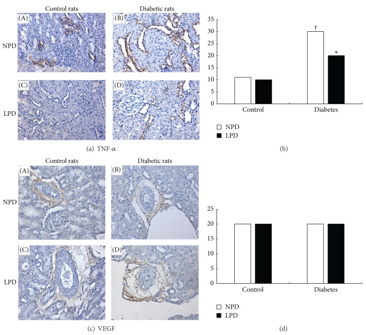
Figure 6.
Effects of LPD on tumor necrosis factor α (TNF-α) and vascular endothelial growth factor (VEGF) expressions in glomeruli and tubulointerstitium. (a) Immunohistochemical staining of TNF-α. TNF-α mainly expressed in the tubulointerstitial area either in control or diabetic rats. As compared with control rats ((A), (C)), diabetic rats ((B), (D)) expressed more abundant TNF-α (the brown color) in this area. LPD only significantly reduced TNF-α expression in diabetic rats (d) but not in control rats (C). (b) Quantitative scores of TNF-α expression in tubulointerstitium. (c) Immunohistobiochemical staining of VEGF. VEGF occasionally expressed around some arterioles. Diabetic rats ((B), (D)) expressed comparable VEGF as control rats ((A), (C)). LPD exerted no effects on VEGF expressions either in control (C) or diabetic rats (D). (d) Quantitative scores of TNF-α expression around arterioles. In (B) and (D), NPD is shown as (□) and LPD is shown as (■).† P < 0.05 compared with C-NPD group. *P < 0.05 is compared with D-NPD group.

