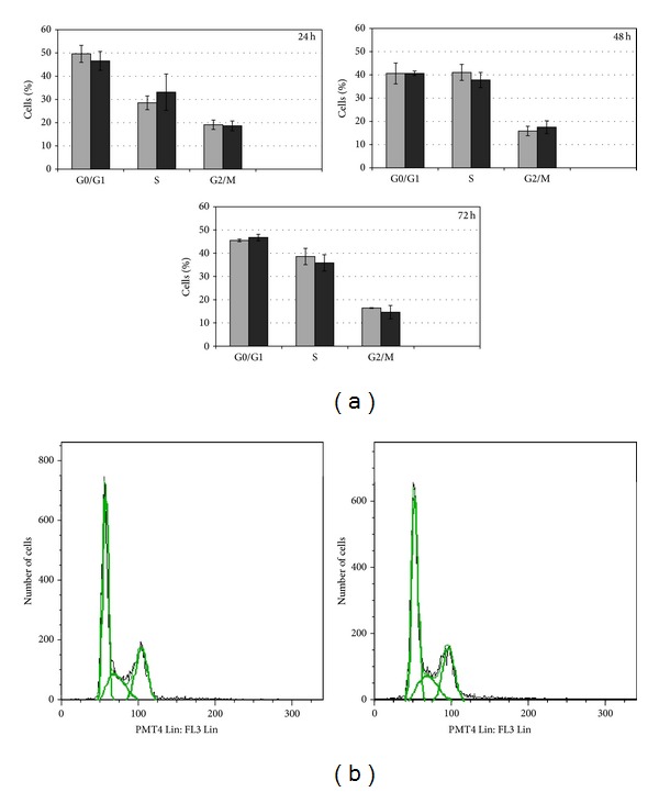
Figure 1.

Effects of CdCl2 on cell cycle distribution of HepG2 cells. HepG2 cells were cultured in the presence of 10 μM Cd concentration for different time points (24, 48, and 72 h). (a) The distribution of the cell population in the different cell cycle phases of treated cells (black bar) is always comparable to controls (grey bar) at all tested time points. In (b) an example of histogram obtained by flow cytometer analysis is displayed; the DNA content (x axis) is plotted against the cell number (y axis). Cells from three independent experiments were analyzed.
