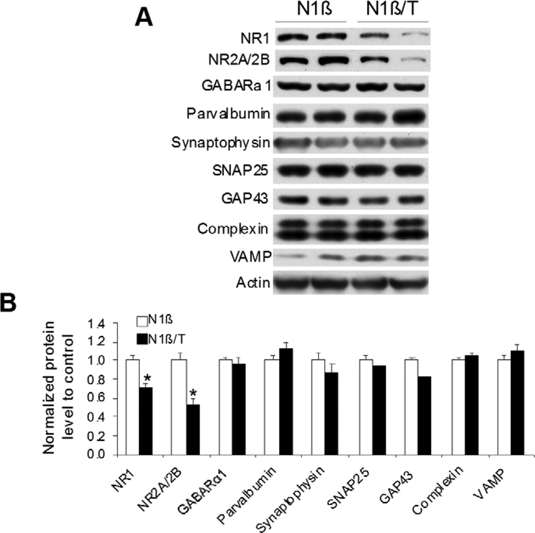
Figure 7. Reduced levels of NMDA receptor proteins in Tg-N1β/T mice.
(A) Protein extracts from 3-month-old mouse hippocampus were examined by Western blotting using antibodies specific to the indicated proteins. Two pairs of mice were used for protein extractions. (B) Bar graphs are shown with the indicated protein levels normalized to actin (n=3 per genotype; *:p<0.05; Student’s t-test.).

