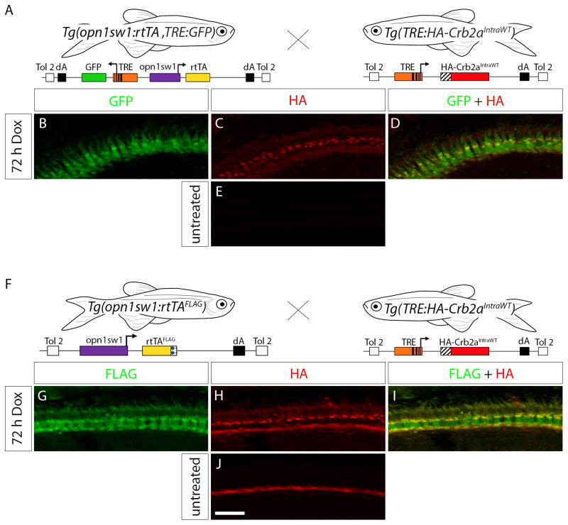
Figure 6. Transactivation in the Tg(TRE:HA-Crb2aIntraWT) line.
(A) Diagram indicating the cross of Tg(opn1sw1:rtTA, TRE:GFP) to Tg(TRE:HA-Crb2aIntraWT) for double-transgenic embryo production. (B–D) Confocal z-projection of a retinal section from a 6 dpf Tg(opn1sw1:rtTA, TRE:GFP; TRE:HA-Crb2aIntraWT) larva treated with Dox for 72 h and labeled with anti-HA antibody. (B) GFP fluorescence (green) and (C) anti-HA immunofluorescence (red) indicate that HA-Crb2aIntraWT is expressed in UV cones (D, merge, yellow). (E) Confocal z-projection of an untreated 6 dpf Tg(opn1sw1:rtTA, TRE:GFP; TRE:HA-Crb2aIntraWT) retinal section labeled with anti-HA antibody. (F) Diagram indicating the cross of Tg(opn1sw1:rtTAflag) to Tg(TRE:HA-Crb2aIntraWT) for double-transgenic embryo production. (G–I) Confocal z-projection of a retinal section from a 6 dpf Tg(opn1sw1:rtTAflag; TRE:HA-Crb2aIntraWT) larva treated with Dox for 72 h and labeled with anti-FLAG and anti-HA antibodies. (G) Anti-FLAG immunofluorescence (green) and (H) anti-HA immunofluorescence (red) indicate HA-Crb2aIntraWT is expressed in the UV cones (I, merge, yellow). (J) Confocal z-projection of an untreated retinal section from a 6 dpf Tg(opn1sw1:rtTAflag; TRE:HA-Crb2aIntraWT) larva labeled with anti-HA antibody. Scale bar (J), 10 μm.

