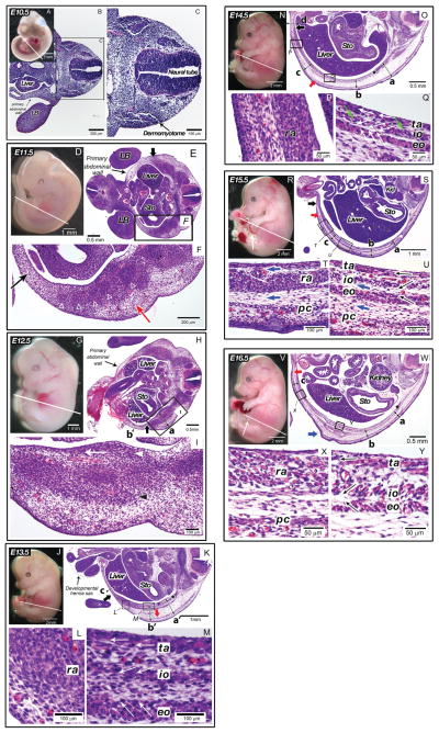
Fig. 2.
Histological characteristics of secondary abdominal wall development in the mouse (E10.5–E16.5). In all figures, the right and left sides correspond to the dorsal and ventral orientations of embryos, respectively. (A–C) E10.5 mouse embryo (CS 14). The primary abdominal wall was closed and myoblasts has not reached to the body wall. (A) Whole mount E10.5 mouse embryo. White line indicates plane of sections shown in B and C. (B) Transverse section of the embryo at low magnification. The limb bud (LB) and liver were seen in this view. The primary abdominal wall is indicated by a black arrow. (C) High magnification of area of interest seen in B. The dermomyotome is indicated by a black arrow. (D–F) E11.5 mouse embryo. (CS 16) Myoblasts have migrated half the distance to the midline, but individual muscles were not seen. (D) Whole mount E11.5 mouse embryo. White line indicates plane of section in E and F. (E) Transverse section of E11.5 mouse embryo. The limb buds (LB) and liver and stomach (Sto) were seen in this view. The primary abdominal wall is indicated by a thin black arrow. The leading edge of myoblast migration is indicated by a thick black arrow at the top of this figure. (F) High magnification view of E11.5 mouse embryo abdominal wall. White open triangles indicate areas of condensations that likely represent early ribs. Black and red arrows indicate mesenchyme cells in the primary and secondary body wall, respectively. (G–I) E12.5 mouse embryo. (CS 18) Myoblasts began organizing into separate muscle layers. (G) Whole mount E12.5 mouse embryo. White line indicates plane of section in H and I. (H) Transverse section of the E12.5 mouse embryo. The liver and stomach (Sto) were seen in this view. The primary abdominal wall is indicated by a thin black arrow. The distance of myoblast migration is indicated by a thick black arrow at the bottom of this figure. The thickness of the body wall was measured: a, 750 μm; b, 180 μm. The distance between the external oblique and the dermis was 280 μm. (I) High magnification view of E12.5 mouse embryo abdominal wall. White arrowhead indicates the inner layer of muscle demonstrating unidirectional orientation (white arrow), black arrowhead indicates the outer layer of muscle. (J–M) E13.5 mouse embryo (CS 20). All muscle groups were visualized but the rectus has not segregated from the other muscle groups and migration to the midline was not yet complete. (J) Whole mount E13.5 mouse embryo. White line indicates plane of section in K–M. (K) Transverse section through E13.5 mouse embryo. The liver and stomach (Sto) were seen as is the developmental hernia sac at the umbilicus. Black star indicates rib. Red arrow indicates ventral most point of the panniculus carnosus, black arrow indicates ventral most point of migrating myoblasts. The thickness of the body wall was measured: a′, 667 μm; b′, 628 μm; c, 82 μm. The length of the connective tissues between the external oblique and dermis was 470 μm at both positions of a′ and b′. (L) High magnification view of the rectus abdominis (ra). (M) High magnification view of the transversus abdominis (ta), internal (io) and external obliques (eo). White arrows demonstrate the directional orientation of myoblasts within individual muscle layers. (N–Q) E14.5 mouse embryo (CS 23). The rectus has segregated from the remaining layers, migration to the midline was 90% completed. (N) Whole mount E14.5 mouse embryo. White line indicates plane of section in O–Q. (O) Transverse section through E14.5 mouse embryo. Red arrow indicates ventral most point of the panniculus carnosus, black arrow indicates ventral most point of migrating myoblasts. Black star indicates the rib. The thickness of the body wall was measured: a, 640 μm; b, 366 μm; c, 400 μm; d, 126 μm. (P) High magnification view of the rectus abdominis (ra). (Q) High magnification view of the transversus abdominis (ta), internal (io), and external obliques (eo). White arrows demonstrate directional orientation of myoblasts within individual muscle layers. Green arrows indicate myotubes. (R–U) E15.5 mouse embryo. Migration of secondary abdominal wall structures to the midline was complete. (R) Whole mount E15.5 mouse embryo. White line indicates plane of section. White arrow indicates developmental hernia sac at the umbilicus. White line indicates plane of section in S–U. (S) Transverse section through E15.5 mouse embryo. The liver, stomach (Sto) and kidney (Kid) were seen in this view. Red arrow indicates ventral most point of panniculus carnosus, black arrow indicates ventral most point of the rectus abdominis. White star indicates the rib. The thickness of the body wall was measured: a, 258 μm; b, 350 μm; c, 305 μm. (T) High magnification view of the rectus abdominis (ra) and panniculus carnosus (pc). Myotubes were now abundant within these muscles. The directions of connective tissues between muscles are shown in blue arrows. (U) High magnification view of the transversus abdominis (ta), internal oblique (io), and external oblique (eo), and panniculus carnosus (pc). Myotubes were abundant within these muscles with the unidirectional orientation (black arrows). The directions of connective tissues between muscles are shown in blue arrows. (V–Y) E16.5 mouse embryo. The closure of the developmental hernia at the umbilicus has occurred complete. (V) Whole mount E16.5 mouse embryo. White line indicates plane of section in W–Y. White arrow indicates the umbilicus after reduction of developmental hernia sac. (W) Transverse section through E16.5 mouse embryo. The liver, stomach (Sto) and kidney were seen in this view. Red arrow indicates ventral most point of panniculus carnosus, blue arrow indicates condensation of a tissue rostral to the hind limb. Black star indicates the rib. The thickness of the body wall was measured: a, 312 μm; b, 351 μm; c, 291 μm. (X) High magnification view of the rectus abdominis (ra) and panniculus carnosus (pc). Unidirectional orientation within the myotubes was seen in the rectus abdominis. (Y) High magnification view of transversus abdominis (ta), internal oblique (io), external oblique (eo). Unidirectional orientation of myotubes was seen in each layer (arrows).

