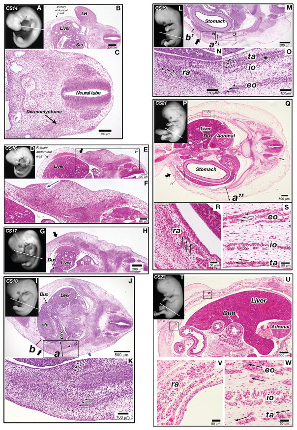
Fig. 3.
Histological staging of secondary human abdominal wall development CS 14–23. In all figures, the right and left sides correspond to the dorsal and ventral orientations of embryos, respectively. (A–C) Human embryo at CS 14 (day 32). The primary abdominal wall was closed and myoblasts has not reached to the body wall. (A) Whole mount photo of a human embryo at CS 14 demonstrating plane of section (white line). (B) Transverse section at the level of the white line in A. Dermomyotome was formed. The limb buds (LB), liver and stomach (Sto) were seen in this view. The primary abdominal wall is indicated by the black arrow. (C) High magnification image demonstrating the dermomyotome and the neural tube. (D–F) Human embryo at CS 16 (day 37). Myoblasts have migrated quarter the distance to the midline. (D) Whole mount photo of a human embryo at CS 16 demonstrating plane of section (white line). (E) Transverse section at the level of the white line in D. The liver was seen in this view. The primary abdominal wall is indicated by the thin black arrow. Thick black arrow at the top of this figure indicates ventral most point of migrating myoblasts. (F) High magnification image of area of interest in image E. An arrow indicates the condensed lateral plate mesoderm. (G and H) Human embryo at CS 17 (day 41). Myoblasts have migrated half the distance to the midline. (G) Whole mount photo of a human embryo at CS 17 demonstrating plane of section (white line). (H) Low magnification photomicrograph of the CS 17 human embryo at the level of a white line in G. The duodenum (Duo) and liver were seen in this view. Black arrow indicates the ventral most point of myoblast migration. Open white arrowheads mark condensations that are likely early ribs. (I–K) Human embryo at CS 18 (day 44). Myoblasts began organizing into separate muscle layers. (I) Whole mount photo of a human embryo at CS 18 demonstrating plane of section (white line). (J) Low magnification photomicrograph of the CS 18 human embryo at the level of the white line in I. The liver, stomach (Sto), duodenum (Duo) and pancreas (panc) were all seen in this view. Myoblasts were organizing into individual layers (area inside the box). Extent of ventral migration indicated by a black arrow. A black star indicates an early rib. The thickness of the body wall measurements were 500 μm (a) and b: 260 μm (b). The connective tissue in the dorsal regions comprises half the thickness of the secondary abdominal wall (blue arrow). (K) High magnification image from image J. Black arrows indicate early directional organization of migrating myoblasts. (L–O) Human embryo at CS 19 (day 47.5). All muscle groups were separated including the rectus abdominis. (L) Whole mount photo of a human embryo at CS 19 demonstrating plane of section (white line). (M) Low magnification photomicrograph at the level of the white line in L. The stomach was seen in this view. Abdominal wall demonstrating that the myoblasts have organized into four distinct muscle layers. Extent of ventral migration indicated by black arrow. The body wall thicknesses were 500 μm (a′) and 260 μm (b′). (N) High magnification view of the rectus muscle at CS 19 demonstrates unidirectional orientation of myoblasts (black arrows). (O) High magnification view of the external oblique (eo), internal oblique (io) transversus abdominis (ta). Black arrows indicate unidirectional organization of myoblasts. The starred structure between the transversus and internal oblique is likely an early nerve. (P–S) Human embryo at CS 21 (day 52). Myoblasts have reached the ventral midline and myotubes were now present and oriented uniformly within all muscle groups. (P) Whole mount photo of a human embryo at CS 21 demonstrating plane of section (white line). (Q) Low magnification photomicrograph at the level of the white line in P. The liver, stomach, adrenal gland and duodenum (Dou) were all seen in this view. Myoblast migration to the midline was complete and the ventral most boundary of the rectus abdominis is indicated by a black arrow. The distance between the external oblique and the dermis was 700 μm and the total thickness of the body wall is 1200 μm at the position a″. (R) High magnification view of the rectus abdominis (ra). Black arrows indicate directional organization of myotubes. (S) High magnification view of the external oblique (eo), internal oblique (io) transversus abdominis (ta). Black arrows indicate directional organization of myotubes which were abundant at this stage. (T–W) Human embryo at CS 23 (day 56.5). Distinct layers were formed in the rectus abdominis and myotube formation became more prominent in all muscles. (T) Whole mount photo of a human embryo at CS 23 demonstrating a plane of section (white line). (U) Low magnification photomicrograph at the level of the white line in T. Liver, kidney and duodenum (Dou) are all seen in this view. (V) High magnification view of the rectus abdominis (ra) in which myotubes are abundantly seen. (W) High magnification view of the external oblique (eo), internal oblique (io) transversus abdominis (ta). Again myotubes were seen in this view. Black arrows represent orientation of the connective tissues between muscle layers.

