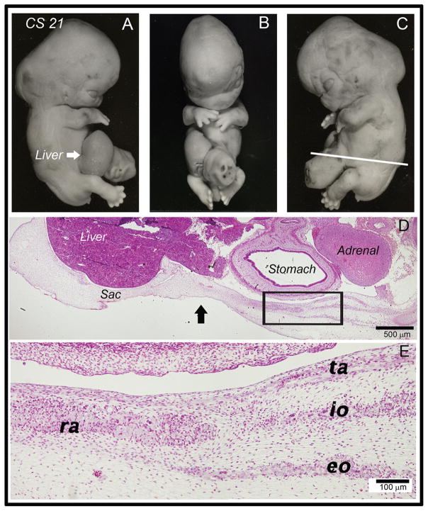
Fig. 4.
Histological analysis of a human omphalocele at CS 21. (A–C) Whole mount photographs of a CS 21 human embryo with omphalocele. Rupture of the sac has occurred on the right and the liver was protruding (white arrow). White line in (C) indicates plane of section for (D) and (E). (D) Low magnification view of a section through this specimen at the level of the white line. The right and left sides correspond to the dorsal and ventral orientations of embryos, respectively. The liver, adrenal gland and stomach are all seen in this view. A large portion of the liver is seen within the developmental hernia sac (area to the left of the black arrow). Four distinct layers of muscle developed. (E) High magnification view of the rectus abdominis (ra), external oblique (eo), internal oblique (io) transversus abdominis (ta). Myoblasts have not yet differentiated into myotubes. There was a lack of unidirectional orientation of myoblasts in the rectus abdominis, external oblique, and internal oblique layers.

