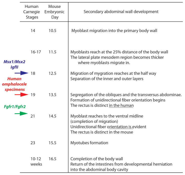
Fig. 6.
Comparative staging of secondary abdominal wall development in the human and mouse. Histological correlation between mouse embryonic day and human CS from Figs. 3 and 4 is presented. Red arrow indicates the approximate timing of arrest in secondary abdominal wall development of the two omphalocele specimens in Figs. 5 and 6. Blue arrow indicates the timing of arrest in Msx1/Msx2 and IGF-II mutants based on published data in which muscle layers stop migration at the half way and no muscle layers are segregated, resulting giant omphalocele (Eggenschwiler et al., 1997). Green arrow indicates the timing of arrest in Fgfr1/Fgfr2 mutants in which the rectus, obliques and transversus abdominis are all present and fails to reach the midline, resulting small omphalocele (Nichol et al., 2011).

