Fig. 5.
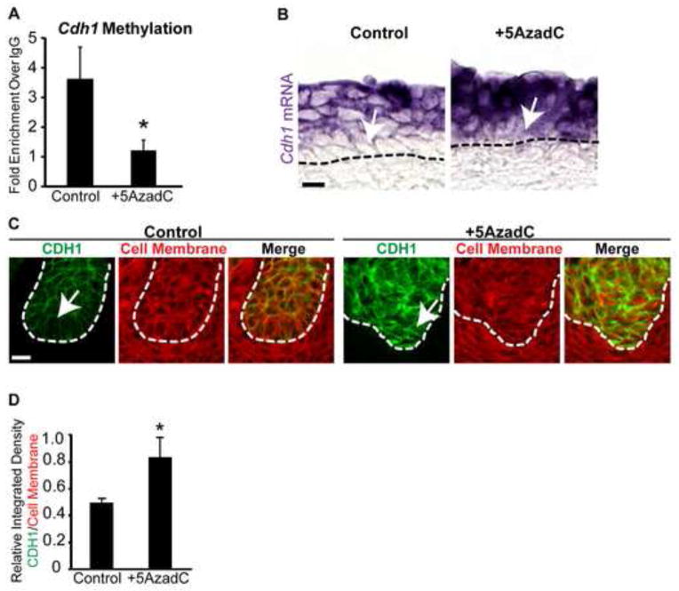
DNA methylation is required for appropriate E-cadherin mRNA and protein expression in basal epithelium of UGS explant cultures. (A) MeDIP-QPCR was used to quantify E-cadherin (Cdh1) methylation in epithelium from 14 dpc UGSs cultured for 4 days in media containing 5α-dihydrotestosterone (DHT, 10 nM) alone or DHT plus the DNA methylation inhibitor 5-aza-2′-deoxycytidine (5AzadC, 5 μM). Female 14 dpc UGSs cultured for 7days in media containing DHT (10 nM) alone or DHT plus 5AzadC were cut into 5 μm sagittal sections and (B) stained by ISH to visualize Cdh1 mRNA expression (purple) and (C) IHC to visualize CDH1 expression (green) and cell membranes (wheat germ agglutinin, red). (D) Quantification of relative integrated density for CDH1 (green) versus cell membrane (wheat germ agglutinin, red) within the basal-most layer in control and 5AzadC treated UGSs. Results are presented as mean ± s.e.m, n ≥ 3 litter independent UGS sections and tissues per group. Asterisks indicate a significant difference from control (p < 0.05). A dotted line indicates the interface between epithelium and mesenchyme, as determined by cell morphology. Arrows indicate cell layers with altered E-cadherin mRNA and protein abundance. Scalebar 10 μm.

