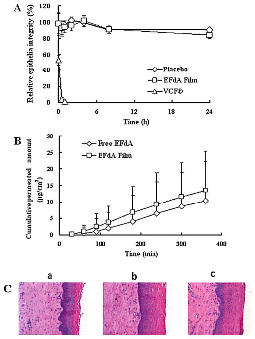
Fig. 6.
(A) Relative HEC-1A epithelial monolayer integrity of placebo and EFdA-loaded films as a function of time; (B) the cumulative amount of EFdA permeated through the human ectocervical tissues versus time. (C) Comparison of morphology of human ectocervical tissues: (a) EFdA film post-exposure; (b) Free EFdA post-exposure; (c) Pre-exposure. Each point represents mean ± SD (n = 4).

