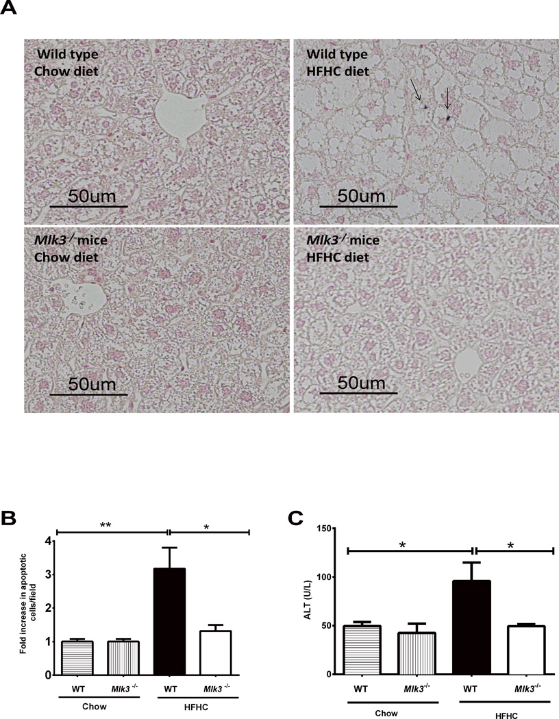
Figure 3. Mlk3−/− mice are protected against high fat high carbohydrate (HFHC) diet-induced hepatic apoptosis, and increased alanine aminotransferase (ALT).
(A) Hepatocytes apoptosis was quantified in paraffin-embedded hepatic tissue of 16 week–fed HFHC diet and chow wild type (WT) and mixed lineage kinase (Mlk3)−/− mice by labeling DNA strand breaks by the terminal deoxynucleotidyl transferase-mediated deoxyuridine triphosphate nick-end labeling (TUNEL) assay. Apoptotic nuclei were stained brown (black arrows) and quantified by counting nuclei in 10 random 20 × microscopic fields per animal. (B) Apoptotic nuclei were expressed as fold increase over control chow-fed WT mice, which was arbitrary set at 1. (C) Serum ALT values were measured by photometric absorbance based technique for the WT and Mlk3−/− mice on chow and HFHC diet for 16 weeks. Data represent the mean ± SEM; * p < 0.05 and **p < 0.01.

