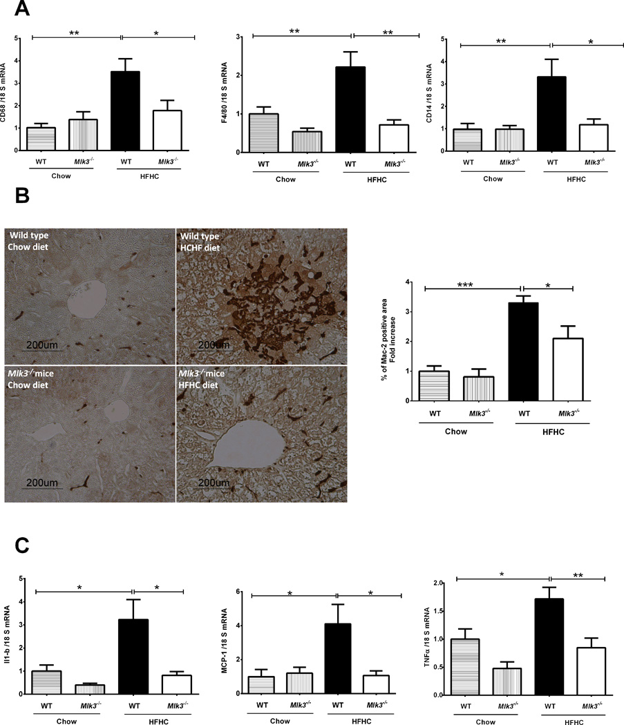
Figure 4. Mlk3−/− mice are protected against high fat high carbohydrate (HFHC) diet-induced hepatic macrophage infiltration and activation.
(A) Total RNA was extracted from the liver tissue of wild type (WT) and mixed lineage kinase (Mlk3)−/− mice on chow and HFHC diet and mRNA expression of macrophage markers cluster of differentiation (CD)68, F4/80 and CD14 were evaluated by real-time polymerase chain reaction (PCR). Fold induction was determined after normalization to 18S mRNA expression. (B) Quantification of macrophage infiltration in the WT and Mlk3−/− mice on chow and HFHC diet was done by immunohistochemistry using macrophage galactose-specific lectin (Mac-2) antibody. Mac-2 immunohistochemical staining was quantified in ten random 20 × microscopic fields per animal by morphometry (KS 400 software, Carl Zeiss). Mac-2 positive area in the liver tissue sections was normalized to control chow-fed WT animals, which was arbitrary set at 1. (C) mRNA expression of cytokines and chemokines related to macrophage activation including interleukin-1 beta (IL-1β), monocyte chemotactic protein-1 (MCP-1), and tumor necrosis factor alpha (TNFα), was assessed by real time PCR, fold induction was determined after normalization to 18S mRNA expression. Data represent the mean ± SEM; *p < 0.05, ** p < 0.01 and ***p < 0.001.

