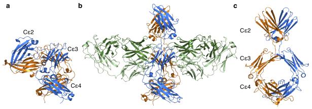
Figure 1. Bent and extended structures adopted by IgE-Fc.
(a) The bent structure of free IgE-Fc, with the (Cε2)2 domain pair making contact with the Cε3-4 domains. IgE-FcA is shown in blue, and IgE-FcB in orange. (b) The structure of IgE-Fc bound symmetrically by two aεFab molecules (shown with heavy chains in dark green, and light chains in light green). (c) The extended conformation of IgE-Fc as seen in the complex (rotated 90° relative to b).

