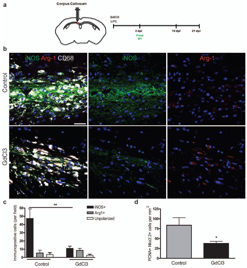
Figure 4. Selective depletion of M1 microglia/macrophages in a demyelinated lesion in the CNS impairs OPC proliferation.
(a) Gadolinium chloride (GdCl3) was injected into corpus callosum lesions at the onset of demyelination (0 dpl) prior to the peak in M1 polarization at 3 dpl. (b) Representative images of control or GdCl3-injected lesions at 3 dpl with immunostaining against iNOS (green), Arg-1 (red), and CD68 (white). Scale bar, 25 μm. (c) Numbers of iNOS+ M1, Arg-1+ M2, or unpolarized (iNOS-, Arg-1-) cells per field ± s.e.m. in control and GdCl3-injected lesions at 3 dpl. One way ANOVA and Newman-Keuls post-test, **p<0.01 (n=5 mice, df=29). (d) Density of PCNA+ Nkx2.2+ proliferating OPCs per mm2 in control and GdCl3-injected lesions ± s.e.m. at 3 dpl. P=0.0496, 2-tailed Student’s t-test (n=5 mice, df=8).

