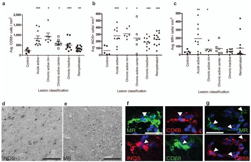
Figure 7. M2 microglia/macrophage densities are increased in acute active and the rim of chronic active multiple sclerosis lesions.
(a) Total CD68+ microglia/macrophages/mm2 were significantly increased in acute active (P=0.001), chronic active rim (P=0.0156) and centre (P=0.0156), chronic inactive (P=0.001) and remyelinated lesions (P=0.002). (b) iNOS+ M1 microglia/macrophages/mm2 were increased in acute active (P=0.001), chronic active rim (P=0.0156) and centre (P=0.0313), chronic inactive (P=0.0005), and remyelinated lesions (P<0.0001). (c) MR+ M2 microglia/macrophages/mm2 were increased in acute active lesions (P=0.0068) and the rim of chronic active lesions (P=0.0156). Mann-Whitney test. n for each lesion type indicated in Supplementary Table 1 online. (d) Representative image of MS lesion with iNOS immunolabeling. Scale bar, 100 μm. (e) Representative image of MS lesion with MR immunolabeling. Scale bar, 100 μm. (f) Co-localization of MR (top) and iNOS (bottom) with microglia/macrophage marker CD68 (arrowheads). Scale bar, 25 μm. (g) MR+ CD68+ M2 cells (arrows) were sometimes associated with blood vessels. Scale bar, 25 μm.

