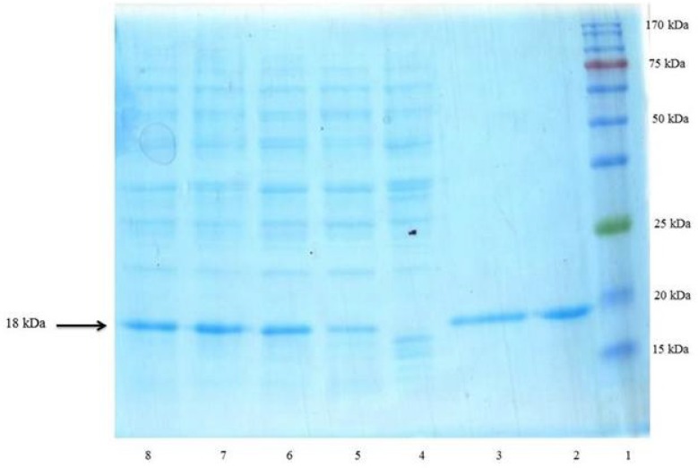
Figure 4.
SDS–PAGE analysis of rhINF-β periplasmic expression in E. coli. Lane 1: Chromatine prestained protein ladder (SinaClon, Tehran, Iran), Lane 2: rhINF-β as positive control (ZiferonTM, Zist Daru Danesh, Tehran, Iran), Lane 3: rhINF-β as positive control (Betaseron®, Bayer HealthCare, Germany), Cell lysates were analyzed before IPTG addition (Lane 4) and 1-4 h after addition of 0.2 mM IPTG (Lanes 5-8). The additional band with molecular weight of 18 kDa in periplasmic fraction after induction corresponds to rhINF-β. The arrow indicates position of rhINF-β

