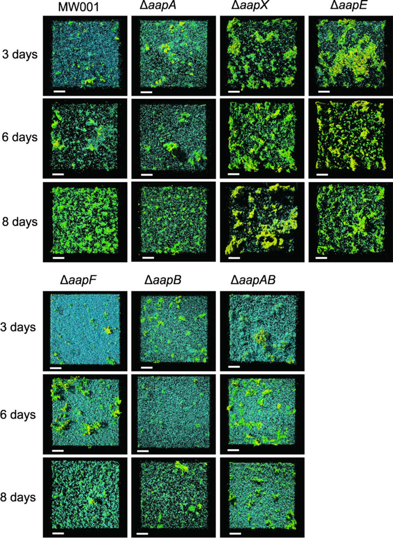
Figure 7.
Static biofilm formation of all aap deletion mutants in comparison to the wild type MW001 analyzed by confocal laser scanning microscopy. Cells were incubated for three, six and eight days in small petri dishes at 75°C in special metal boxes to prevent evaporation of medium. The cells were visualized using DAPI (blue channel) and EPS was stained using the two fluorescently labeled lectins ConA and IB4. The green channel represents ConA which binds to glucose and mannose residues, whereas IB4 binds to α-galactosyl residues (yellow channel).. Overlay of all three channels are shown. Scale bars are 40 µm.

