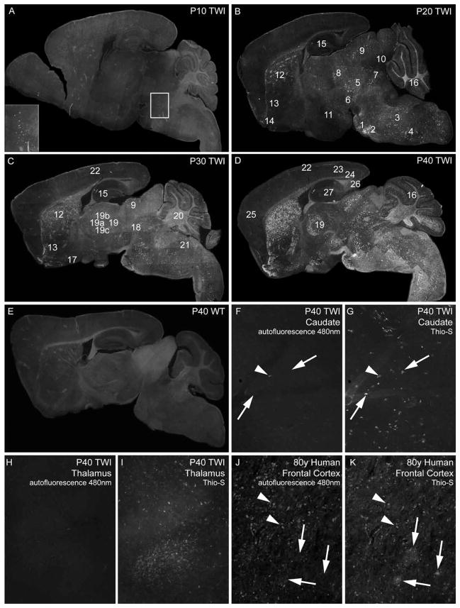
Figure 1. Distribution of Thioflavin-S reactive material in the Twitcher brain.
A–E) Composite mapping of thioflavin-S (Thio-S) reactive deposits at 10 (A), 20 (B), 30 (C) and 40 (D) days of age in the Twitcher (TWI) brain. E shows the composite image of thioflavin-S staining of a 40 day-old wild type (WT) brain. Insert in A marks the pontine region (pons). 1, pontine grey area; 2, superior olivary complex; 3, medulla; 4, spinal nucleus of the trigeminal nerve; 5, midbrain; 6, substantia nigra; 7, reticular nucleus; 8, dorsal lateral geniculate nucleus; 9, superior colliculus; 10, inferior colliculus; 11, hypothalamus; 12, caudate putamen; 13, nucleus accumbens; 14, olfactory tubercle; 15, hippocampus; 16, cerebellar internal granular layer; 17, hypothalamic pre-optic area; 18, superior red nucleus; 19, thalamus; 19a, lateral dorsal nucleus; 19b, anteroventral nucleus; 19c, reticular nucleus; 20, cerebellar arbor vitae; 21, vestibular nuclei; 22, cortex layer 1; 23, cortex layers 3 and 4; 24, cortex layer 6b; 25, frontal cortex; 26, subiculum and 27, dentate gyrus. Composite images were taken using a 5× objective and each contain 60–70 individual images. F–K) Unstained (F, H) and thioflavin-S stained (G, H) TWI caudate and thalamus tissue imaged under 480nm filter. Scarce autofluorescent material (arrowhead) seen in TWI compared to abundant thioflavin-S reactive inclusions (arrows). Positive control of autofluorescent lipofucsin accumulations (arrowheads) compared to thioflavin-S reactive deposits (arrows) shown in aged human frontal cortex (J, K). Images in (F–K) taken with 10× objective.

