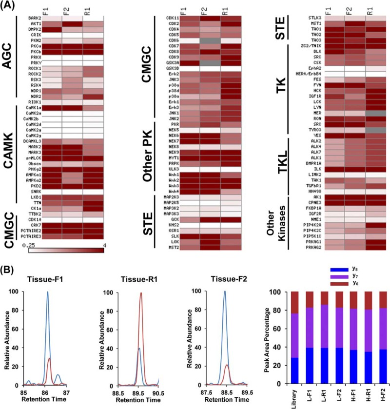
Fig. 7.
MRM-based global kinome profiling revealed differential expression of kinases in lung tumor and adjacent normal lung tissue. A, A heatmap showing the differential expression of kinases from tumor and adjacent normal lung tissue based on Rtumor/normal ratio in two forward and one reverse labeling reactions. Dark red and white boxes designate those kinases that are up-regulated in tumor tissue and normal tissue, respectively, as indicated by the scale bar. B, Quantitative results by MRM assay for peptide DLK#PSNLLINTTCDLK from MAPK3 kinase: (Left and Middle) Extracted ion chromatograms for three transitions monitored for light-labeled (Red) and heavy-labeled (Blue) peptides in both forward and reverse labeling reaction; (Right) the consistent distribution of the peak area observed for each monitored transition from light- and heavy- labeled peptides in both forward and reverse labeling reaction along with the theoretical distribution derived from MS/MS spectra stored in MRM kinome library.

