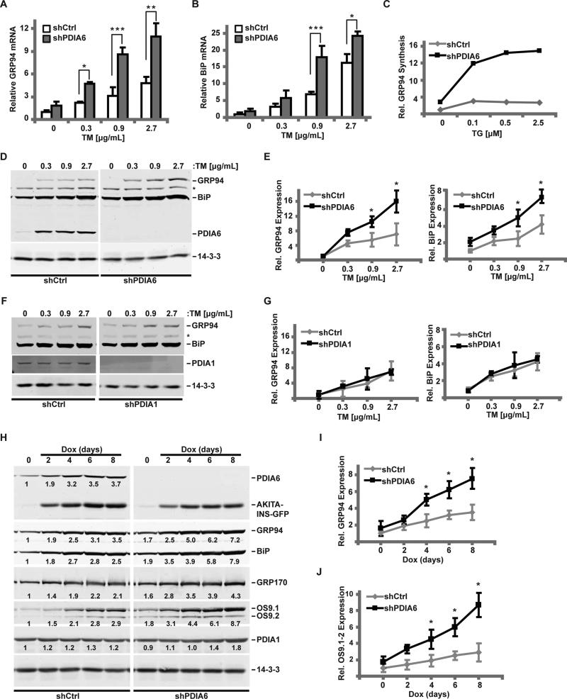
Figure 1. Loss of PDIA6 causes an augmented unfolded protein response.
(A-B) 3T3 cells expressing a non-targeting (shCtrl) or a PDIA6-targeting shRNA (clone 1) were treated with the indicated dose of TM for 6hrs. GRP94 (A) and BiP (B) mRNA levels were detected by RT-qPCR, normalized to β-actin, and their expression in shCtrl cells without stress was set at 1. Plots are means±SD, n=3. Significant differences between shPDIA6 and shCtrl conditions are indicated by asterisks. (*, p ≤ 0.05; **, p ≤ 0.01; ***, p ≤ 0.001 (student t test)).
(C) shCtrl or shPDIA6 3T3 cells were exposed to the indicated dose of TG for 6hrs and labeled with [35S]Met/Cys for 30 min. Relative GRP94 synthesis was determined by immunoprecipitation and normalized to untreated shCtrl cells.
(D) shCtrl or shPDIA6 (clone 1) 3T3 cells were treated with varying concentrations of TM for 16 hrs and levels of GRP94, BiP or PDIA6 were determined with αKDEL antibody. 14-3-3 was used as a loading control. *, an unknown KDEL-positive protein whose expression is unchanged.
(E) Quantitation of GRP94 and BiP levels from panel D. Values are means±SD relative to DMSO-treated shCtrl cells (*, p ≤ 0.05; n=3).
(F-G) shCtrl or shPDIA1 3T3 cells were treated with TM and analysed as in panels D-E.
(H-J) Akita-INS cells were transduced with shCtrl or shPDIA6 (clone 1) lentiviruses and Akita insulin expression was induced with 2μg/mL doxycycline (Dox). The relative levels of indicated proteins were measured by immunoblotting and normalized to the loading control, 14-3-3 (numbers below bands). The induction of GRP94 (I) or OS9.1/OS9.2 (J) was quantified as in panels D-E.
See also Figs. S1-S2.

