Fig. 5.
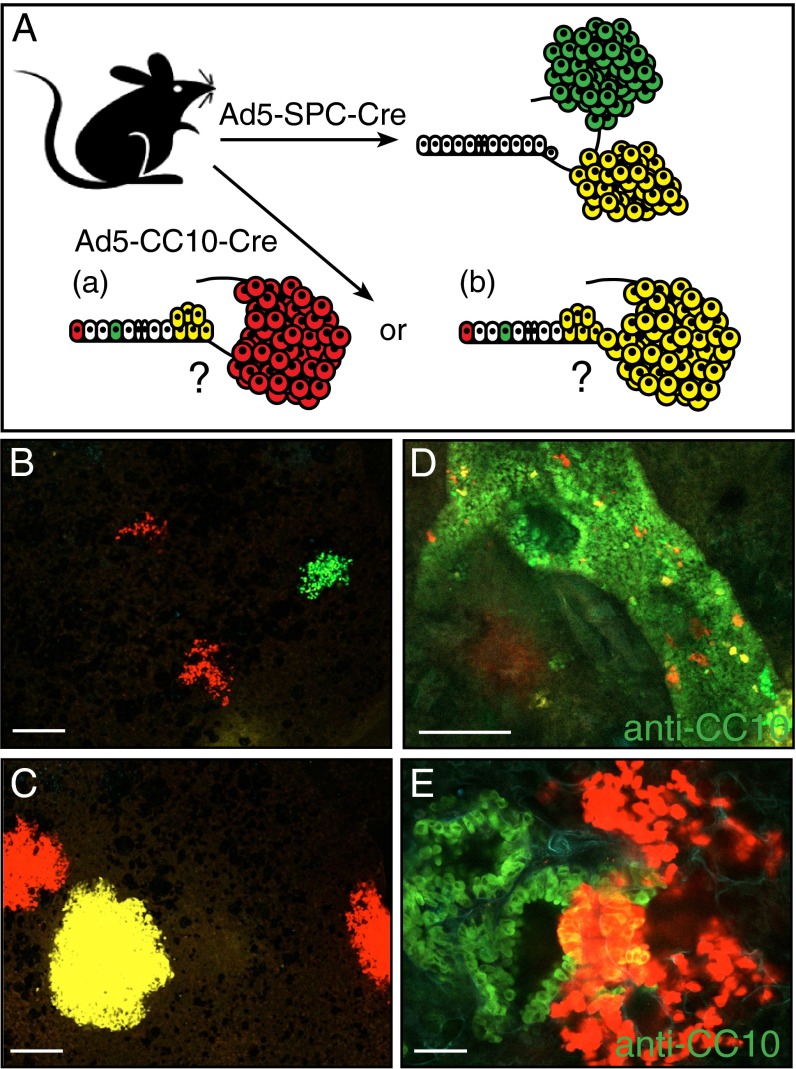
Clonal outgrowth of CC10+ and SPC+ cells following K-RasG12D activation. (A) Schematic representation of the genetic strategy to trace CC10- and SPC-expressing cells in K-RasG12D lung lesions. Regarding the lesions observed in K-RasLSL-G12D/+ mice following Ad5–CC10–Cre infection, (a) do the lesions in the alveoli arise from CC10-expressing cells present in the alveoli, or (b) do the lesions in the alveoli arise from transformed cells present at the BADJ? (B and C) Confocal imaging of multiple areas of alveolar hyperplasia (B) and AAH/adenomas (C) present in the lungs of K-RasLSL-G12D/+;R26R-Confetti animals following Ad5–SPC–Cre infection. Each lesion is marked uniformly by a single Confetti color. (D and E) Confocal imaging of CC10-stained lung tissue sections from a K-RasLSL-G12D/+;R26R-Confetti mouse following Ad5–CC10–Cre infection. (D) Single Confetti+ switched cells resident on the bronchiolar wall stained positive with anti-CC10 immunofluorescence staining. This immunofluorescence staining pattern indicates that not all K-RasG12D–expressing (Confetti+) switched cells are capable of further expansion/proliferation. (E) Areas of clonal hyperplasia (red Confetti color) located at the terminal bronchiole contain cells that coexpress the Clara cell marker, CC10 (yellow). (Scale bar in B–D, 100 μM; E, 500 μM.)

