Fig. 1.
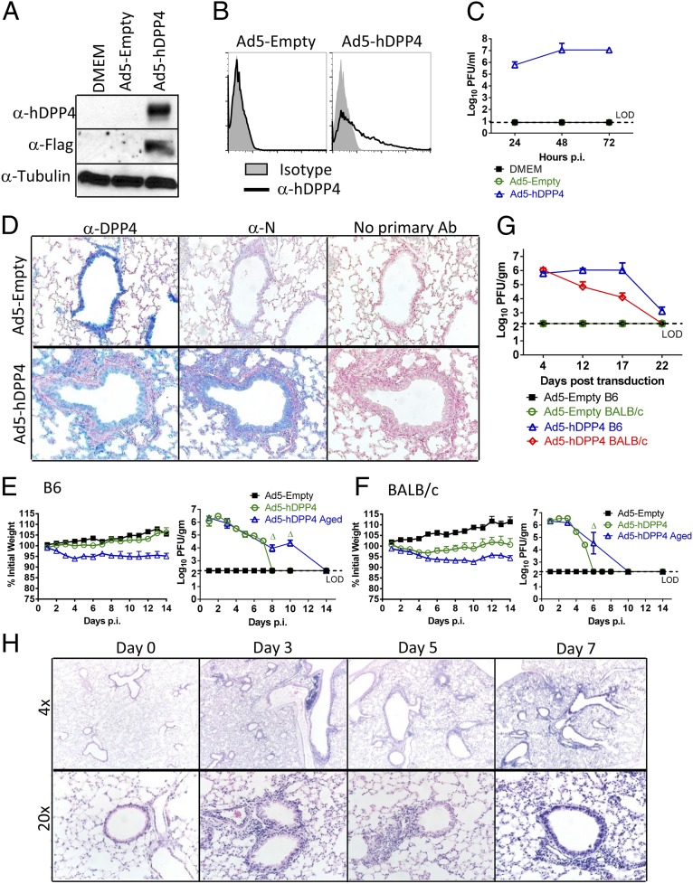
Development of mice susceptible to MERS-CoV infection. To assess hDPP4 expression (A) and surface localization (B), MLE15 cells were transduced with Ad5-hDPP4 or Ad5-Empty at an MOI of 20 at 37 °C for 4 h. hDDP4 expression was monitored by Western blot assay (A) or flow cytometry (B). (C) Ad5-hDPP4–transduced cells were infected with MERS-CoV at an MOI of 1 at 48 h posttransduction, and virus titers were determined by plaque assay. Five days after transduction with 2.5 × 10⁸ pfu of Ad5-hDPP4 or Ad5-Empty in 75 μL of DMEM intranasally, mice were intranasally infected with 1 × 10⁵ pfu of MERS-CoV in 50 μL of DMEM. (D) Lungs were harvested from BALB/c mice at day 3 after MERS-CoV infection, fixed in zinc formalin, and embedded in paraffin. Sections were stained with an anti-hDPP4 or with an anti–MERS-CoV nucleocapsid antibody (blue signal). (Original magnification, 20×.) (E and F) Weight changes in 6- to 12-wk-old (young) and 18- to 22-mo-old (aged) B6 (E) and BALB/c (F) mice were monitored daily. For B6 mice, n = 8 in Ad5-Empty group; 12 in Ad5-hDPP4 group; 8 in Ad5-hDPP4 aged group. For BALB/c mice, n = 8 in Ad5-Empty group; 12 in Ad5-hDPP4 group; 8 in Ad5-hDPP4 aged group. To obtain virus titers, lungs were homogenized at the indicated time points and titered on Vero 81 cells. Titers are expressed as pfu/g tissue (n = 4–8 mice per group per time point). Data are representative of two independent experiments. Δ, P < 0.05 when Ad5-hDPP4 aged were compared with Ad5-hDPP4 and Ad4-Empty. (G) To evaluate the length of time that Ad5-hDPP4–transduced mice could be infected with MERS-CoV, Ad5-hDPP4–transduced mice were infected with MERS-CoV at the indicated times. Lungs were harvested for titers at 2 d.p.i. (n = 4 mice per group per time point. (H) Lungs from B6 mice were removed at the indicated time points p.i., fixed in zinc formalin, and embedded in paraffin. Sections were stained with hematoxylin/eosin.

