Fig. 4.
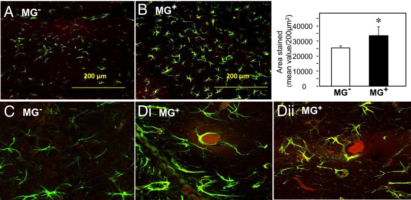
Oral MG+ promotes brain gliosis and AGE deposits. Confocal microscopy of coronal hippocampal sections from (A) MG+ and (B) MG− mice (n = 8/group) immunostained for glia cells (Magnification, 20×), and (C) MG− and (D, i and ii) MG+ for both glia and AGEs, using anti-GFAP and anti-AGE, as primary antibodies and Alexa Fluor-488 (green) and Fluor-594 (red), respectively, as secondary antibodies (magnification, 200×). Bar graph shows the quantification of GFAP staining. (Scale bar, 200 µm.) *P < 0.05, MG+ vs. MG− mice.

