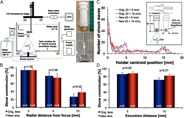
Fig. 4.
In vitro stone comminution. (A) Schematic of the in vitro stone comminution experimental setup with tube holder depicted and pictured at right. (B) stone comminution results in the tube holder positioned at three radial distances in the lithotripter focal plane (r = 0, 5, and 10 mm). (C) Illustration of simulated respiratory motion (Inset) and average motion histograms corresponding to (D) stone comminution results at two excursion distances (D = 5 and 15 mm).

