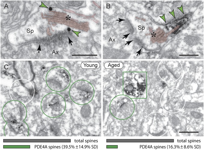
Fig. 4.
PDE4A is lost from dendritic spines in aged monkey dlPFC. (A and B) In young dlPFC, PDE4A (green arrowheads) is localized next to the SA (asterisk); shown are immunogold and immunoperoxidase, respectively. (Scale bars: 200 nm.) (C) PDE4A is widely expressed in layer III spines in young, but not in aged monkey dlPFC (green circles, PDE4A spines; white circles, unlabeled spines; green rectangle, PDE4A dendrite). (Scale bar: 0.5 μm.) The graphs illustrate percentage of PDE4A spines in 10-y vs. 25-y monkey dlPFC (Upper), averaged from 46-μm2 electron microscopic fields (20 fields from five tissue blocks per animal; SI Materials and Methods) of layer III. Young (Left): PDE4A spines/46 μm2 = 5.95 (± 2.13 SD). Aged (Right): PDE4A spines/46 μm2 = 2.05 (± 1.24 SD).

