Fig. 1.
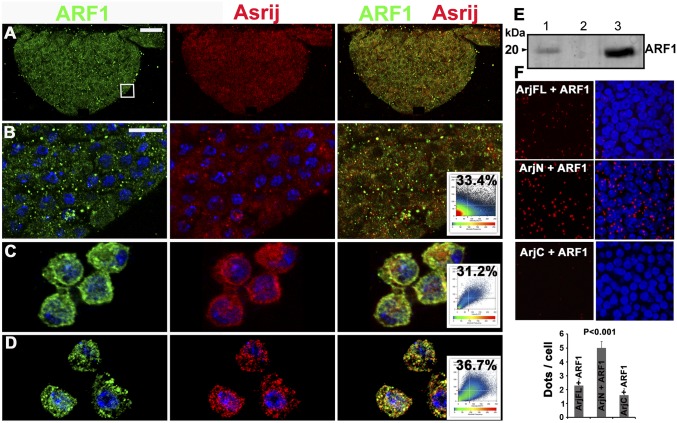
ARF1 interacts with the pan hemocyte marker Asrij. (A–D) ARF1 (green) and Asrij (red) colocalization in the lymph gland (A, and magnified boxed region in B), hemocytes in circulation (C), and S2R+ cells (D). Colocalization plots are as indicated. (E) Coimmunoprecipitation (co-IP) of Asrij and ARF1 from S2R+ protein extracts with anti-Asrij antibodies. Immunoblot was probed with anti-ARF1 antibody. (Lane 1) Input control (10% of the total protein). (Lane 2) IP with preimmune serum. (Lane 3) IP with anti-Asrij antibodies. (F) In situ proximity ligation assay on wild-type lymph glands using antibodies against ARF1 and Asrij full length (ArjFL) or ArjN or ArjC. Graph shows PLA signal (red dots/cell). n = 10. Nuclei were stained with DAPI (blue). [Scale bar, 20 μm (A) and 5 μm (B–D and F).]

