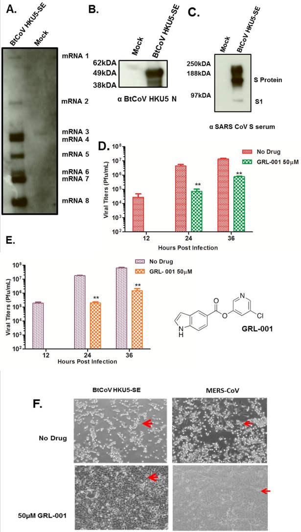
FIG 2 .
Subgenomic mRNA and protein expression in BtCoV HKU5-SE and the 3C-like protease inhibitor study. (A) Northern blot showing subgenomic mRNA expression in BtCoV HKU5-SE-infected cells. (B and C) Western blots showing expression of structural proteins N (B) and S (C) from BtCoV HKU5-SE-infected cell lysates stained with polyclonal serum as indicated below the images. (D and E) Viral titers from cells pretreated with 50 µM of GRL-001 and infected with BtCoV HKU5-SE (D) and MERS-CoV (E) at an MOI of 0.1 PFU/ml. Drug treatment was continued after infection, and virus-containing supernatants were sampled in triplicate. Error bars indicate SD. Asterisks indicate statistical significance (P < 0.05, Student t test). The structure of GRL-001 is shown to the right of panel E. (F) Bright-field images of cells infected with BtCoV HKU5-SE or MERS-CoV at an MOI of 0.1, showing strong cytopathic effects with no drug treatment (top, red arrows) and intact monolayers with 50 µM GRL-001 treatment (bottom, red arrows) at 36 h p.i..

