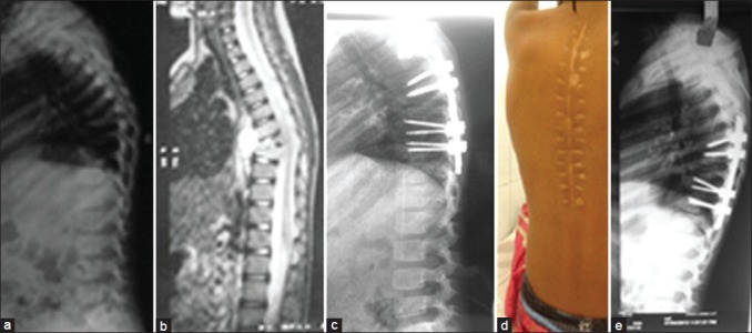
Figure 5.

A 5 year female with (a) kyphus deformity (b) preoperative X-ray and (c) magnetic resonance imaging after 6 months of anti tubercular treatment and (d) postoperative X-ray showing kyphus correction using pedicle screws (e) healed posterior midline incision (e) 2 years followup x-rays of the same patient, the anterior vertebral body height has increased on followup
