
Figure 2C.
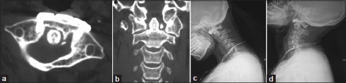
(a and b) Axial and coronal plane computed tomography at 2 years followup showing satisfactory osseous fusion, the good position of the screws and good reduction of the fracture. The lateral displacement of the lateral masses was improved to 2.0 mm (c and d) dynamic flexion/extension lateral radiographs of cervical spine showing no C1-C2 instability