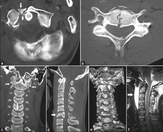
Figure 3A.

(a-e) Preoperative axial plane, coronal plane, sagittal plane computed tomography (CT) scan and three dimensional reconstruction CT shows anterior arc comminution fractures on the right side associated with C6 burst fracture, the height of C6 vertebrae lost half, both of lateral mass displacement are about 6.0 mm (f) T2-weighted images in the sagittal plane show the gap and the signal intensity changes between C1 and C3 level. Prevertebral hematoma is indicated by solid white arrows, the width of prevertebral hematoma is about 9.8 mm in C1-C3 level, dural sac was partly compressed in C6 level, no abnormal spine cord signals
