
Figure 3B.
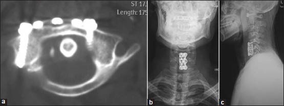
(a-c) At 6 months followup, axial computed tomography and roentgenograph manifested satisfactory osseous fusion and cervical alignment. The patient underwent a corpectomy of C6 and an anterior interbody fusion with a titanium mesh cage spacer and bone grafting of autologous C6 vertebrae