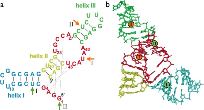
Figure 1.

Diels–Alderase ribozyme. (a) Secondary and tertiary structure interactions in the folded state. Solid lines, secondary structure base pairs; dotted lines, tertiary structure base pairs. Attachment sites of the FRET labels are marked by green (donor dye Cy3 at U6 in construct I and at the 5′ end in construct II) and orange (acceptor dye Cy5 at U42 in construct I and at U30 in construct II) arrows. (b) Three-dimensional structure of the folded state. Color-coding of the secondary structure elements as in panel a. Attachment sites of the FRET labels are indicated by green (Cy3, donor) and red (Cy5, acceptor) spheres. (The figure has been adapted from Figure 1 in ref (9).)
