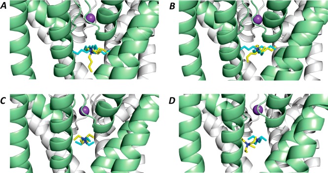
Figure 4.
Lowest energy score docked outputs for TBA (A, B) and TEA (C, D) using Flexidock (A, C) and GOLD (B, D) docked into KcsA (PDB:2BOB for TBA; PDB:2BOC for TEA). In each case the docked small molecule structure is represented by yellow sticks, and the crystal structure coordinates for TBA are represented by blue sticks. The blue TEA structure in panels C and D was made by editing the TBA coordinates to truncate the butyl chains to ethyl chains. In all runs potassium ions occupied the [1] and [3] positions of the selectivity filter; the [3]K+ ion is shown as a purple sphere.

