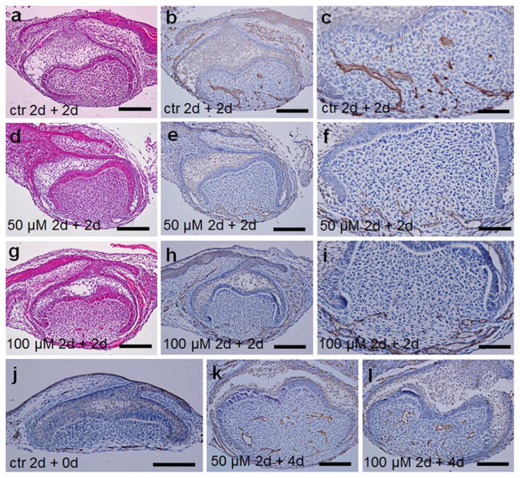
Fig. 4.
The histology, and vascularization of the mouse molar mesenchyme examined by CD31 immunohistochemistry after z-VAD-fmk treatment. E14.0 first lower molars were cultured in vitro in control medium (a–c, j) or in the presence of 50 (d–f, k) or 100 μM (g–i, l) z-VAD-fmk for 2 days, then cultured under mouse kidney capsules for 0 (j), 2 (a–i), or 4 days (k, l). a–c In the control molars, blood vessels appeared in the central part of the dental papilla after 2-day subrenal culture. d–i Blood vessels were only localized in the mandibular portion of the dental mesenchyme and the surrounding tissue in the 50-μM group (d–f) and in the 100-μM group (g–i) after 2-day subrenal culture. j Blood vessels are only detected in the surrounding dental follicle in the molars cultured in vitro in control medium for 2 days. k, lCapillaries are noted not only in the dental follicle but also in the dental papilla after a 2-day culture with the presence of z-VAD-fmk and a following 4-day subrenal culture. a, d, g H&E staining; b, c, e, f, h–l CD31 immunostaining. Bars (a, b, d, e, g, h, j–l) 100 μm, c, f, i 50 μm

