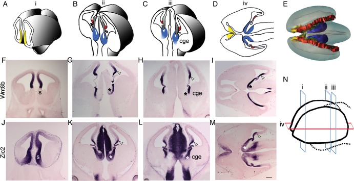
Figure 1.
Medial forebrain structures. (A–C) Schematics of 3 coronal levels of sectioning in the E12.5 mouse brain, corresponding to “i,” “ii,” and “iii” in N, respectively. “i” is at the level of septum (yellow); “ii” and “iii” show the cortical hem (red) and the thalamic eminence (TE; blue). TE is contiguous with the CGE as seen in level “iii.” (D) Schematic depicting the positions of septum, cortical hem, and TE in a horizontal section, corresponding to “iv” in N. The 3 rostro-caudal coronal levels (i, ii, and iii) and 1 horizontal level (iv) of sectioning in the E12.5 whole forebrain are schematized in N. (E) Dorsal view of a 3-D model of E12.5 brain showing the locations of septum (yellow), hem (red), and TE (blue), encircling the choroid plexus (green). Wnt8b is expressed in the medial neuroepithelium of the dorsal telencephalon, including the pallial septum (F), cortical hem (open arrowhead), and also in the ventricular zone of TE (asterisk; G–I). (J–M) Zic2 is expressed in the entire medial forebrain, including the forebrain hem system. CGE, caudal ganglionic eminence; h, hem; s, septum; TE, thalamic eminence. Scale bar: 300 µm.

