Figure 4.
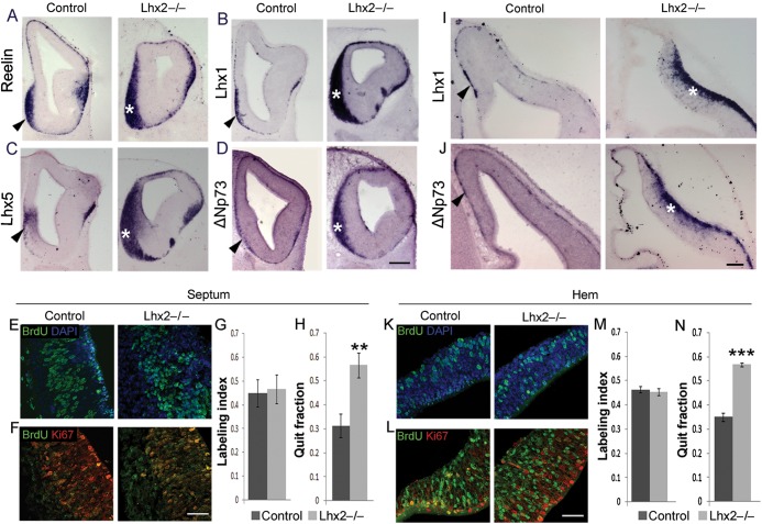
Loss of Lhx2 results in an increase in Cajal–Retzius cells. (A–D) Expression of Reelin (A), Lhx1 (B), Lhx5 (C), and ΔNp73 (D) at E12.5 reveals a considerable increase in the Cajal–Retzius cells accumulated at the septum of Lhx2 mutants (asterisks) compared with control brains (arrowheads). (I and J) Similarly, the mantle of the mutant hem (asterisks) also shows increased Cajal–Retzius cell marker expression compared with controls (arrowheads). (E–H) corresponds to sections and cell counts for the septum and (K–N), for the hem. (E and K) Sections of E11.5 control and Lhx2 mutant brains, after a 2-h BrdU pulse, processed for BrdU and DAPI staining. Labeling indices (BrdU+ cells/total DAPI+ cells) at E11.5 are similar for control and Lhx2 mutant septum (45% and 47%, respectively; E and G) and hem (46% and 45%, respectively; K and M). (F and L) Sections of E12.5 Lhx2 control and mutant brains harvested 1 day after BrdU administration at E11.5 and processed for BrdU–Ki67 double immunohistochemistry. The quit fraction (BrdU+; Ki67− cells/total BrdU+ cells) is significantly greater in the Lhx2 mutant septum (57%; **P < 0.005) and hem (57%; ***P < 0.0005) than in the control septum (31%) and hem (35%). Scale bars: 300 µm (A–D); 50 µm (E, F, K, L); 100 µm (I, J).

