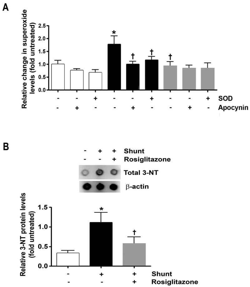
Figure 6.
Superoxide (A) and nitrotyrosine (3-NT) protein levels (B) in lung tissue from normal (white bars) and vehicle (black bars)- and RG-treated (gray bars) shunt lambs. (A) Superoxide levels estimated by electron paramagnetic resonance. Superoxide levels where higher in vehicle-treated shunt lambs than normal lambs, and lower in RG-treated than vehicle-treated lambs. Specificity of the EPR assay for superoxide was confirmed by a reduction in the waveform amplitude with the addition of superoxide dismutase (SOD) to the samples, in vehicle-treated shunt lambs. The addition of apocynin (an NADPH oxidase inhibitor) to the samples resulted in no change in EPR amplitude of normal and RG-treated lambs, but a ∼37% signal reduction in vehicle-treated lambs. (B) Lung tissue 3-NT levels were increased in vehicle-treated shunt lambs compared to normal lambs, and decreased in RG-treated compared to vehicle-treated shunt lambs. Representative Western blots are shown. N=5 for normal group. N=5 for vehicle-treated shunt group N=6 for RG-treated shunt group. Values are mean ± SD. *P<0.05 compared to normal group. †P<0.05 compared to vehicle-treated shunt group.

