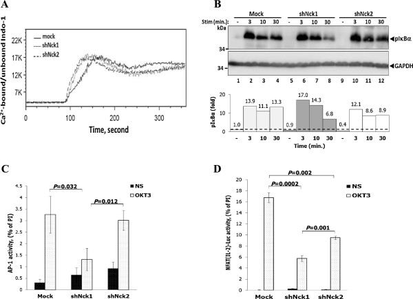
Figure 4.
Dispensable role of Nck protein in TCR-induced Ca2+ mobilization. A) Cells were loaded with Indo-1 AM and baseline was measured for 1 min before stimulation with OKT3 antibody. The ratio of Ca2+-bound and -unbound forms of Indo-1 AM was monitored by flow cytometry. Data are representative from three independent experiments. B) Cells were stimulated with soluble anti-CD3 antibody (1 μg/ml) at various time points. Lysates were immunoblotted with anti-phospho-IκBα (Ser32) antibody. Below, signal intensity was quantified and presented as a ratio of p-IκBα to GAPDH relative to that in unstimulated control cells (Mock), set as 1 (black dashed line). Data are representative of two independent experiments. C) Cells were transiently co-transfected with pAP-1-Luc and control pGL4.7 plasmid. After 20 hr of transfection, cells were left untreated or stimulated with plate-bound monoclonal antibody to CD3, or with a combination of PMA and ionomycin. Cells were then lysed and measured for the luciferase activity. To control for the transfection efficiency, the pAP-1-Luc activity was normalized to the Renilla luciferase activity. Bars represent the mean luciferase activities ± SD from triplicate wells and expressed as percentage of the response to PMA plus ionomycin (PI) and are representative of two independent experiments. D) Each cell population was transiently co-transfected with the pNFAT(IL2)-Luc reporter plasmid plus control pGL4.7 plasmid. After 20 hr of transfection, cells were carried out as described above. Bars represent the mean luciferase activities ± SD and expressed as percentage of the response to PMA plus ionomycin (PI). The results were compared among the groups by using the two-tailed unpaired t test. The p-value less than 0.05 were considered for statistical significance. The data are representative of two independent experiments.

