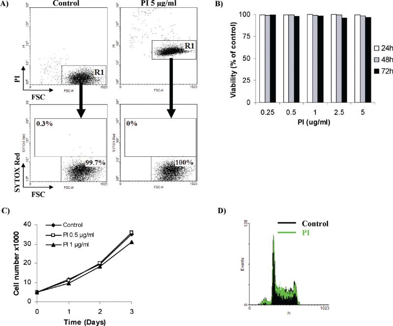
Figure 2.
Continuous exposure to PI has no effect on subsequent exclusion of SYTOX Red. (A) Human promyelocytic leukemia HL60 cells were exposed to increasing concentrations of propidium iodide (PI; 0.25–5 μg/mL) for up 72 h. Cell viability was estimated afterward using spectrally distinct fluorescent probe SYTOX Red. Note that no change in cell viability was assessed by exclusion of SYTOX Red following continuous exposure to PI. Similar results were achieved with SYTOX Green, SYTOX Red, PO-PRO 1, and YO-PRO 1. Results represent the mean of three independent experiments with SD values consistently lower than ± 3. (B) Reults show that the increased fluorescent signal from PI does not imply loss of cell viability. Human promyelocytic leukemia HL60 cells were incubated with or without 5 μg/mL PI for 24 h. Cells were then counterstained with 5 nM SYTOX Red and analyzed by flow cytometry. Note that the R1 subpopulation with elevated PI fluorescence (in the right panel) is not SYTOX Red+ and is thus presumed viable. Results are representative of three independent experiments. (C) Results show that PI does not disturb cell proliferation. HL60 cells were continuously challenged with low doses of PI (0.5 and 1 μg/mL) and the number of viable cells was assessed using the standard Trypan Blue assay during the 3 day study. Note the lack of cell growth retardation. The results represent the mean of at least three independent experiments. Normalized SD values were lower than ± 3. (D) Results show that exposure to PI does not disturb cell proliferation. HL60 cells were continuously challenged with 0.5 μg/mL PI for up to 48 h. Cells were then fixed, and their DNA content profile was analyzed by flow cytometry. Note the lack of any cell cycle disturbances.

