
Figure 5. Model-predicted effects of degradation and sequestration on VEGF patterning.
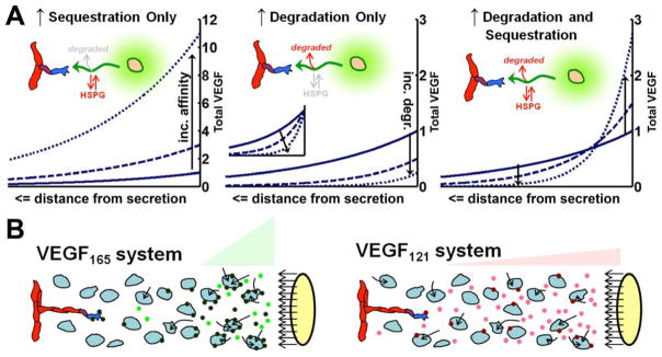
A, Isoform-specific VEGF patterning as seen in vivo relies on isoform-specific differences in both degradation and sequestration (19). The graphs show the spatial distribution of the total VEGF (soluble + sequestered) for three VEGF isoforms (representing increasing matrix binding affinities and/or degradation). All conditions have identical secretion rates. Graphs are scaled to maximum concentration of the lowest affinity isoform (VEGF121 – solid line, identical in each case), except the inset in the middle graph which shows each distribution normalized to its maximum concentration, to show relative steepness. Arrows indicate effect of increasing degradation and/or sequestration. B, In vivo, different rates of uptake of VEGF isoforms by surrounding tissues may account for experimentally observed gradients. We hypothesize that VEGF165 (left) shows greater pericellular accumulation and localization because of dual effects of greater sequestration by HSPGs or cell-surface receptors, and greater degradation (loss from the system) by the resultant cellular internalization. Secretion of VEGF121 (right) has lower binding to cells and lower degradation, and thus increased levels in solution and dispersed spatial gradients. Internalization (arrows) may be due to interstitial cells as well as the endothelium, because both cell types can express VEGF receptors.